Corneal Tear Repair
Corneal lacerations are common in ocular trauma. Repair should occur within 24 hours to reduce the risk of infection, alleviate patient discomfort, and avoid further damage to the eye. The goal for repair is a watertight closure, restoration of normal anatomy, and prevention of high astigmatism or scarring. This activity reviews the evaluation and management of corneal lacerations and highlights the role of the interprofessional team in managing this condition.
Objectives:
- Identify the initial steps for evaluation when evaluating a patient with an eye injury.
- Describe the process for treatment of a corneal laceration.
- Review surgical techniques for repairing a corneal laceration.
- Summarize the postoperative complications that can occur after a corneal laceration repair.
Introduction
Ocular injury is common, with an estimated 24 million people in the United States suffering an eye injury. Injuries to the eye vary in severity, from a small scratch to the cornea (corneal abrasion) to a split in the external structure (globe rupture). Globe rupture can occur in various parts of the eye; one example is a corneal laceration. In a review of 890 eye injuries in Iraq and Afghanistan from 2001 to 2011, 20.7% involved a corneal laceration.
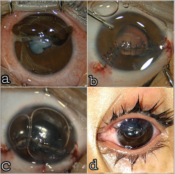
Corneal lacerations vary in size and shape, can be partial or full-thickness, and range from a simple linear pattern to a complex stellate formation. All lacerations require urgent repair to reduce the risk of infection, decrease tissue necrosis, and alleviate patient discomfort. The typical recommendation for a repair is within 24 hours.
The repair of a corneal laceration often requires suturing; however, tissue adhesives or contact lenses can close lacerations less than 2 mm.
The goal of any repair is a watertight closure, restoration of normal anatomy, and limiting the amount of post-operative corneal scarring and astigmatism.
Ocular trauma is an emergency which should be addressed immediately. Injury to cornea is a preventable cause of blindness. A meticulous corneal tear repair is essential for optimal visual outcome.
Principles of corneal tear repair
Place the first suture at limbus when it is involved.It provides anatomic stability to wound edges.
Place suture perpendicular and equidistant to the cut edge at 85-90% depth to ensure optimum tension for layer-to-layer approximation.Equal amount of tissue should be incorporated on each side of the wound.
In shelved/oblique lacerations, sutures to be placed equidistant with respect to the internal aspect of the wound. Apply long tight compressive suture at the periphery.Central sutures should be minimally compressive and short.This results in peripheral flattening and central steepening of the cornea.
Bury suture knots in corneal stroma to lessen post-op inflammation and infection.
Revise any loose or too tight sutures to achieve regular corneal contour. Cornea flattens adjacent to tight sutures and steepens adjacent to loose sutures, hence affecting astigmatism significantly.