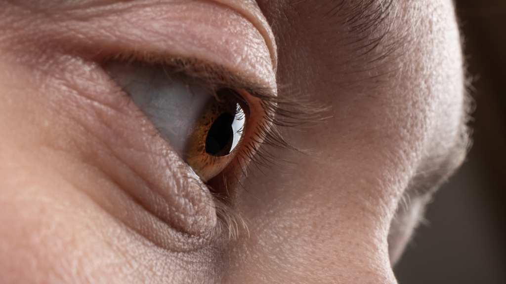
Corneal Tattooing
Tattooing of cosmetically disfiguring corneal scars may be a valuable therapeutic alternative in a distinct group of patients. This group comprises patients in whom reconstructive surgical procedures either will not result in functional improvement or carry the risk of phthisis. Besides this, increasing difficulty in wearing a printed contact lens or a bulbar shell or the reluctant attitude of the patient towards repeated surgery (or enucleation) may be of importance. Out of the numerous modifications reported for corneal tattooing, it is not easy to choose the optimal one.
Introduction
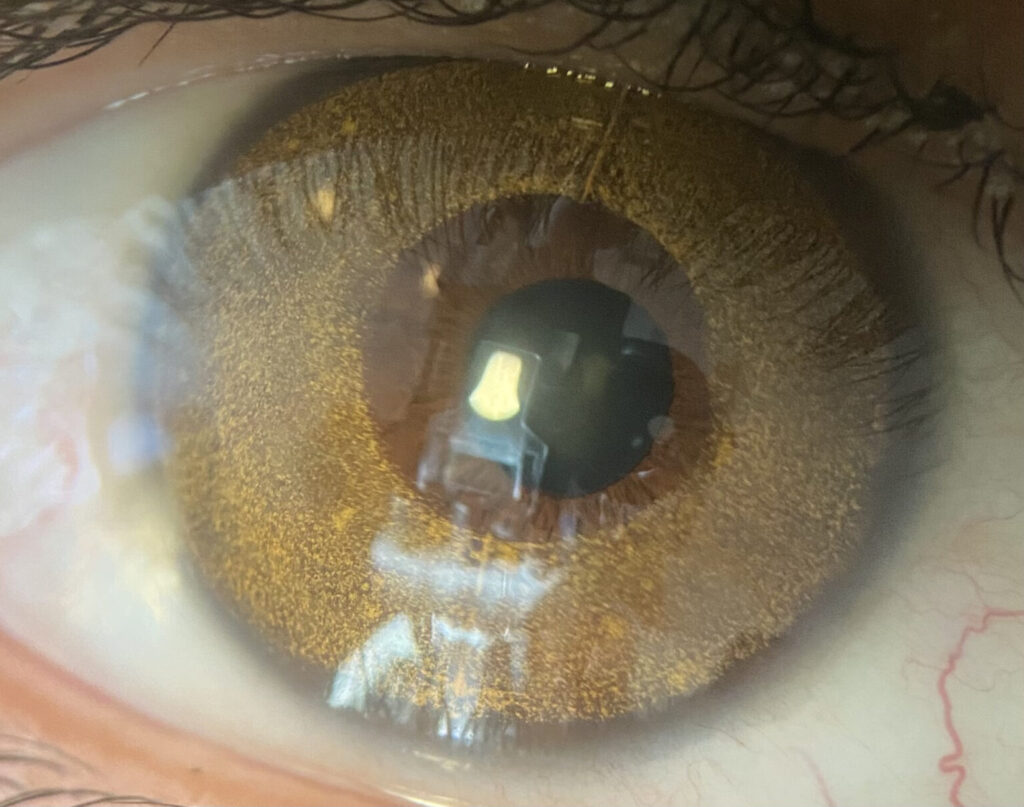
Keratopigmentation or corneal tattooing is a procedure used for centuries to improve the cosmetic appearance of corneal scars and leukomas.
Keratopigmentation has been used to also treat visual symptoms associated with corneal and iris abnormalities. Several techniques have been developed using a variety of ink pigments to achieve the best functional and cosmetic results.
A valuable therapeutic alternative in a select group of patients in whom non-surgical (ex. contact lenses) and reconstructive surgical procedures will not result in functional or cosmetic improvement. Most common indications include corneal scars, leucomas, aniridia, polycoria, traumatic iris defects, and iridodialysis.
Other uses reported in the literature include symptomatic glare or diplopia associated with laser peripheral iridotomies and intractable binocular diplopia.
Techniques
Staining method: Direct application of tattoo ink to anterior surface of cornea. Benefits include fast procedure with uniform dye application. Limitations of this technique include relative impermeance of dye resulting in a high risk of fading.
Impregnation method: Tattoo pigment is directly introduced into the corneal tissue by needle puncture. Benefit is longer retention of pigment but more difficult and time consuming procedure. Irregularly placed pigment deposits can cause light scatter.
Lamellar keratectomy technique: Benefits include longer retention of pigment and lower risk of recurrent corneal erosions. Panda et al. compared a series of patients who underwent conventional (impregnation) tattooing vs lamellar pocket procedure and noted greater pigment density, no episodes of erosion and none of the cases required repeat treatment at 1 year follow-up.
Femto-assisted corneal tattooing (FACT): A femtosecond laser is used to make a superficial corneal pocket into which tattoo ink is injected. The procedure enables the ink to last longer and achieve more uniform cosmetic results. Video of the technique by Dr Keith Walter performed using a femtosecond laser.
Tattoo ink selection: Historically one of the major limitations of corneal tattooing has been the lack of systemic studies demonstrating biocompatibility of pigments and resulting unavailability of reliable commercial products. A variety of sterile ink formulations have been reported in the literature. Micronized mineral pigments are a relatively new pigment that has been shown to produce good cosmetic appearance without an inflammatory response and ocular toxicity.
Complications
Complications of corneal tattooing include corneal infection, inflammation, pain, and risk of inadvertent globe penetration via entry into the anterior chamber. In a case series by Alio et al., keratopigmentation with micronized mineral pigments was performed in 234 eyes of 204 patients and percentage of complications was 12.82% of which the majority was light sensitivity (49%), color fading (19%), and neovascularization (7%).